What is AVM Disease (Arteriovenous Malformation)?
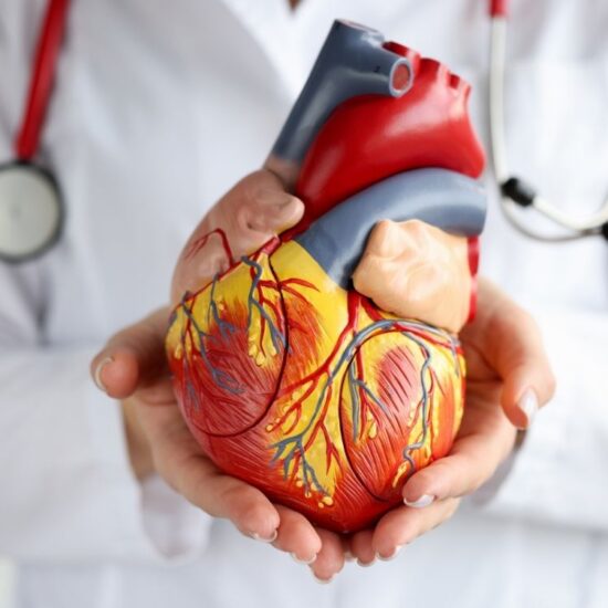
An arteriovenous malformation, also known as an AVM, is a tangle of blood vessels that create irregular connections between arteries and veins. This disrupts blood flow and prevents oxygenation of tissues. An AVM can occur anywhere in the body, including the brain.
Arteries carry oxygen-rich blood from the heart to the brain and other organs. Veins drain oxygen-depleted blood back to the lungs and heart. When an AVM disrupts this critical process, surrounding tissues may not get enough oxygen.
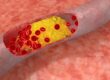
Because the tangled blood vessels in an AVM don’t form properly, they can weaken and burst. If an AVM in the brain bursts, it can cause bleeding in the brain, which can lead to a stroke or brain damage. Bleeding in the brain is called a hemorrhage.
The cause of AVMs is not clear. They rarely run in families.
Once a brain AVM is diagnosed, it can usually be treated to prevent or reduce the risk of complications.
What are the symptoms of AVM disease?
The symptoms of an arteriovenous malformation, also known as an AVM, can vary. Sometimes an AVM doesn’t cause symptoms. An AVM may be found during imaging for another health problem.
Usually, the first symptoms appear after bleeding occurs. In addition to bleeding, symptoms may include:
- A thinking problem that gets worse over time.
- Headaches.
- Nausea and vomiting.
- Seizures.
- Loss of consciousness.
Other possible symptoms include:
- Muscle weakness, for example weakness in the legs.
- Loss of movement and feeling in a part of the body is known as paralysis.
- Loss of coordination that can lead to problems walking.
- Difficulty performing tasks that require planning.
- Back pain.
- Dizziness.
- Vision problems. These may include loss of part of the field of vision, difficulty moving the eyes, or swelling of part of the optic nerve.
- Trouble speaking or understanding language.
- Numbness, tingling, or sudden pain.
- Memory loss or dementia.
- Seeing or hearing things that do not exist is known as hallucinations.
- Clouding of consciousness, confusion.
Children and teenagers may have problems with learning or behavior.
A type of AVM called vein of Galen malformation causes symptoms that appear at birth or shortly after birth. Vein of Galen malformation occurs deep in the brain. Symptoms may include:
- The head becomes larger than normal due to fluid accumulation in the brain.
- Swollen veins on the scalp.
- Seizures.
- Failure to thrive.
- Congestive heart failure.
When should I see a doctor?
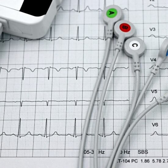
Get medical help if you have any of the symptoms of an AVM, such as headaches, dizziness, vision problems, seizures, and thought changes. Many AVMs are found during testing for another condition, such as a CT scan or MRI.
Causes of AVM Disease
Arteriovenous malformation occurs when arteries and veins connect irregularly. Experts don’t understand why this happens. Certain genetic changes may play a role, but most types don’t usually run in families.
Risk factors
Rarely, a family history of arteriovenous malformations may increase your risk, but most types are not hereditary.
Certain inherited conditions may increase your risk of arteriovenous malformations, including hereditary hemorrhagic telangiectasia, also known as Osler-Weber-Rendu syndrome.
Complications
The most common complications of an arteriovenous malformation are bleeding and seizures. Bleeding can cause brain damage and, if left untreated, can lead to death.
Diagnosis
To diagnose an AVM, also known as an arteriovenous malformation, your health care provider will review your symptoms and give you a physical exam.
Your healthcare provider may listen for a sound called a murmur. A murmur is a whistling sound caused by blood flowing quickly through the arteries and veins of an AVM. It sounds like water flowing through a narrow pipe. A murmur may interfere with your hearing or sleep, or cause emotional distress.
Tests commonly used to diagnose AVM include:
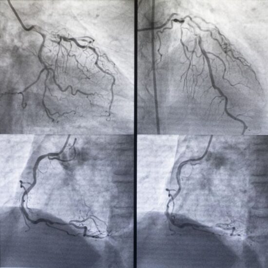
- Cerebral angiography.This test looks for an AVM in the brain. Also called arteriography, this test uses a special dye called contrast medium that is injected into an artery. The dye highlights the blood vessels so they show up better on X-rays.
- CT scan.These scans can help show bleeding. CT scans use X-rays to create images of the head, brain, or spinal cord.
- CT angiography.This test combines a CT scan with an injection of dye to help find a bleeding AVM.
- MR.An MRI uses powerful magnets and radio waves to show detailed images of tissues. An MRI can detect small changes in these tissues.
- Magnetic resonance angiography, also known as MRA.MRA captures the pattern, speed, and distance of blood flow in irregular vessels.
- Transcranial doppler ultrasonography.This test can help diagnose an AVM and tell if the AVM is bleeding. The test uses high-frequency sound waves aimed at the arteries to create an image of blood flow and speed.
Treatment
Treatment for an arteriovenous malformation, also known as an AVM, depends on where it is located, your symptoms, and the risks of treatment. Sometimes an AVM is monitored with regular imaging tests to watch for changes. Other AVMs require treatment. Your healthcare provider may recommend conservative treatment if the AVM has not ruptured and you are not at high risk of bleeding from the AVM.
When deciding whether to treat an arteriovenous malformation, healthcare professionals consider:
- Whether the shopping mall is bleeding or not.
- Whether the AVM is causing symptoms other than bleeding.
- Whether the AVM is in a part of the brain that can be safely treated.
- Other features of the shopping mall, such as its size.
Medicines
Medications can help manage symptoms associated with an arteriovenous malformation, such as seizures, headaches, and back pain.
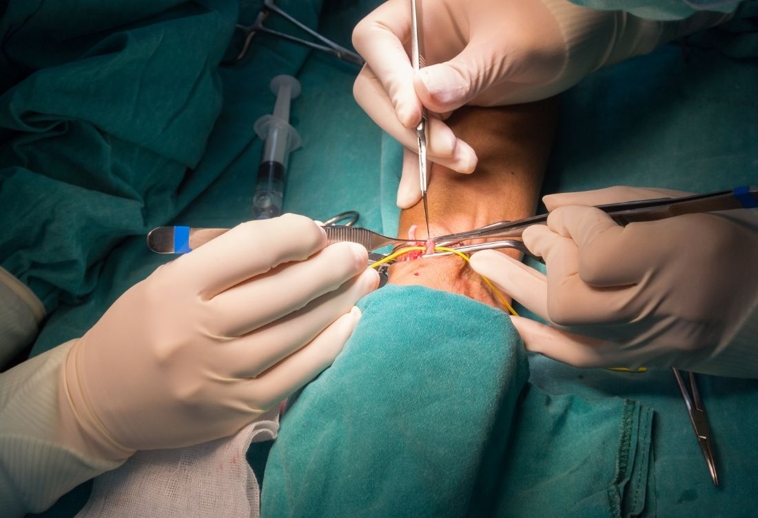
Operation
The main treatment for AVM is surgery. Surgery can completely remove the arteriovenous malformation. This treatment may be recommended if the risk of bleeding is high. Surgery is usually an option if the AVM is in an area where the risk of damage to brain tissue is low.
Endovascular embolization is a type of surgery that involves passing a catheter through the arteries into the arteriovenous malformation. A substance is then injected to seal off parts of the AVM to reduce blood flow. This may be done before brain surgery or radiosurgery to help reduce the risk of complications.
Sometimes stereotactic radiosurgery is used to treat an AVM. The treatment uses intense, highly focused beams of radiation to damage blood vessels. This helps stop blood flow to the AVM.
You and your healthcare team will discuss whether to treat your AVM and weigh the possible benefits against the risks.
To follow
After treatment for an arteriovenous malformation, you may need regular follow-up visits with your healthcare team. You may also need more imaging tests to make sure the AVM was treated successfully and the malformation has not returned. If your AVM is being monitored, you will also need regular imaging tests and follow-up visits with your healthcare team.